At the start of using neuroimaging to try and understand mental health problems, the focus was on a specific area of the brain that might be different. As the methods have become more sophisticated, the ability to look at how different areas of the brain are linked into functional networks has developed.
Whilst damage to a specific part of the brain does lead to a characteristic pattern of behavioural change, one of the most notable features of our brain is just how connected each bit of the brain is to other parts of the brain. No task is completed by just using one part of the brain, and the same area can be involved in different networks for completing different tasks. Additionally, damage to one part of the network can be compensated by changes within the network, or by recruiting extra parts of the brain.
Trying to unpick this complexity is a key task of current neuroimaging research. As MRI scanning is not detailed enough to see how actual neurons connect together, researchers try and estimate functional links between different areas by seeing if the BOLD response (a proxy for some neural activity, but not all, see Swettenham et al, 2013) in one area is statistically associated with another area.
Changes to a series of different functional networks have been proposed to underlie both the susceptibility to depression (often called trait factors and see my previous blog for more on this) and/or actually having depression (state factors). Gathering together different studies to get enough subjects to try and work out if these theoretical networks are indeed associated with depression is one aim of functional MRI meta-analyses.
Methods
Researchers from the UK tried to combine all the studies they could find that had used fMRI in depression and had published enough data to allow the findings to be combined. They split the studies they included into 2 groups
- cross-sectional studies: comparing people with current depression to controls (34 papers, total participants =1,165)
- treatment studies: measuring changes from before treatment to afterwards. (6 studies, total participants = 105). 4 of these studies included a control group.
And they excluded studies where there was no difference between the groups (n=4).
The studies included did not get participants to do the same task in the scanner. This means that the results are a bit tricky to interpret, but if the same networks in the brain are affected by having depression, despite what the person is doing in the scanner, then it provides some evidence that these networks are important.
The researchers used two different statistical methods to combine these studies and compared the results. The results overlapped for some findings, but not for others limiting the conclusions that can be drawn from this study. They were keen to include as much data as possible (to increase power), but that was at the expense of how confident we can be in how specific the results are to depression.
Results
- Widespread areas of the brain were found to be different in depression
- These included areas in all the lobes of the brain and some sub-cortical areas
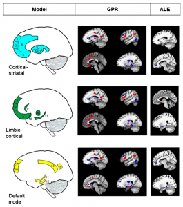
- The researchers linked these findings to three different theoretical models of how brain networks may change in depression
- They found some support for all three, but not enough to favour one model over another
- Getting better from depression was associated with changes in a much smaller range of areas of the brain
- But these were completely different with the two different methods of meta-analysis

Researchers tried to match the results to existing models of depression (click image to view full size)
Conclusions
The researchers hedged their bets:
a meta-analytic description of the neurobiology of depression from fMRI both provides support for previous models, but also suggests some important differences
but they emphasised the big variation in what is currently classified as depression eg it includes ‘melancholia, psychotic depression and post-natal depression’, and therefore questioned whether ‘a single model of depression can be applied’.
Summary
- This study is interesting in trying to combine the results of many different tasks to find out if there is evidence of an underlying disturbance of one specific network in the brain.
- Unfortunately there were too many factors influencing the results for any clear conclusions to be drawn.
- But this study does suggest that a model of depression that relies on changes to only one functional network is too simplistic.
- A more sophisticated breakdown of different symptoms/types of depression is going to be needed for us to get to the bottom of what might be underlying the condition.
Links
Graham, J., et al., Meta-analytic evidence for neuroimaging models of depression: State or trait? Journal of Affective Disorders (2013), http://dx.doi.org/10.1016/j.jad.2013.07.002i [PubMed abstract]
Swettenham JB, Muthukumaraswamy SD and Singh KD (2013) BOLD responses in human primary visual cortex are insensitive to substantial changes in neural activity. Front. Hum. Neurosci. 7:76. doi: 10.3389/fnhum.2013.00076 [Abstract]


Trying to understand brain networks associated with depression is hampered by how variable the condition is: A… http://t.co/if1LOLfMJU
@Mental_Elf 1 condition – or a final common pathway of a number of processes, most as yet not well categorised?
@andrewwatson28 describes #neuroimaging models of #depression by summarising a recent meta-analysis in J-Affect-Dis http://t.co/zKWEgE0Drm
@Mental_Elf deftly summarized #neuroimaging models of #depression 2013 meta-analysis in J-Affect-Dis http://t.co/BDTAXecQRX @andrewwatson28
Still a neurological enigma; “Trying to understand brain networks associated with depression…” http://t.co/w44BvWdLQA
We’ve another top #neuroimaging blog today from @andrewwatson28 This time about #depression http://t.co/zKWEgE0Drm
Don’t miss: Trying to understand brain networks associated w/ depression is hampered by how variable the condition is http://t.co/zKWEgE0Drm
Trying to understand the brain networks associated with depression is hampered by how variable the condition is: http://t.co/SZBKQjrzx0
Mental Elf: Trying to understand brain networks associated with depression is hampered by how variable the… http://t.co/pqmwlt3fxH
In the news: Trying to understand brain networks associated with depression is hampered by variability of condition http://t.co/xLHfDiOu8C
depression is to vague a concept: neuroimaging demos variables in brain activity. http://t.co/mUfWVRoPQn
This makes sense: #Depression is complex. I would think different types are slightly different neurologically…. http://t.co/szBifAR0Z5
Brain changes – I sometimes think about for example hunger, I’m guessing there are changes in the brain when we are hungry importantly this doesn’t indicate we need treatment and possible medication, we need to eat!! I think its the reasons behind the depression that need addressing.