

Does a mouse think like a human? Does a cat? Does a macaque monkey? These are fascinating questions to ask on a philosophical level, but they are also of immense practical importance.
Current regulations on drug development mean that animal research plays a huge role in deciding what substances might be safe and beneficial to humans. This has lead to great success in understanding many aspects of how the body works, and using that to develop treatments.
But is the nervous system different? If it contains the main adaptations that mean humans can do many more complex things than other animals, can working out how an animal’s brain works reap the same rewards? Many drug companies have spent a lot of money on compounds that worked very well on mouse behaviours, but did not have the desired effect in humans. This has contributed to some of them pulling out of neuroscience research altogether. It means that finding ways to better understand how similar/different animals’ brains are from each other can offer both the hope of better treatments for humans, and fewer animals used to find those treatments.
Using imaging to do this allows for more humane studies of animals close to us the evolutionary tree. It also offers the opportunity for speculation about the areas of the brain that ‘make us human’. Whilst it is impossible that there is one area of the brain that contains the essence of humanity, the differences between species offers vital clues as to how the brain functions.
Researchers from Oxford have been combining data from humans and macaque monkeys across a series of studies, and the one we are focusing on today is on a part of the frontal lobe (see figure 1).
The ventral, inferior frontal cortex (vIFC) has been linked with a range of functions, but seems to be heavily involved in modelling the potential consequences of actions and selecting appropriate behaviours for the situation. It includes Broca’s area, long recognised as part of the system that understands and produces speech.
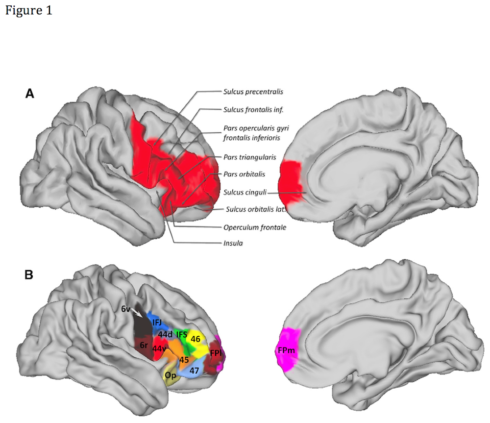
Figure 1 – Click on the image to see a bigger version
Methods
The study had three goals:
- Split up the human vIFC based on where each sub-region connects to anatomically
- See what the pattern of functional connectivity is for these areas
- Compare this pattern to macaque monkeys
In order to do this the researchers scanned 25 healthy humans using MRI. They used two types of MRI scanning: examining the white matter connections in the brain (anatomical connectivity) and functional MRI at rest (functional connectivity). They also scanned 25 macaque monkeys using fMRI while they were anaesthetised.
The researchers used some very sophisticated analysis methods to meet their goals. Every tiny area (a 5x5x5mm voxel) in the vIFC was examined for its connection patterns to the rest of the brain. These connection patterns were then compared to each other and the voxels that had similar pattern were grouped together. Finally these groups were fitted back onto the anatomy of the brain. Figure 1 shows the subdivisions found.
A different method was used to subdivide the monkey’s putative equivalent brain areas. This was done by using already published maps subdividing the macaque brain. These maps are made by identifying areas that have similar patterns of cells, not anatomical connections. This has been done with humans as well (Broadman areas) and the areas described overlap substantially with the imaging findings in this study, but by no means exactly.
Finally the functional connectivity was compared from region to region to see if the human and monkey regions of the brain match up in how they connect to the rest of the brain.
Results
There was both substantial overlap between human and monkey brain function, and key differences. The detail for each area is fascinating and if your neuroanatomy is up to it I’d highly recommend looking at the paper’s figures, e.g. figure 2 for two of the subdivisions of the motor part of the vIFC
- In areas associated with using visual information for motor control and planning the humans and macaques were very similar
- Some human areas implicated in language showed very similar patterns to equivalent monkey areas, despite macaques not having sophisticated language skills.
- This provides support for the idea that there is not a specific/separate language network in humans. Language processing probably ‘built upon existing mechanisms for sensory –motor mapping, sequential motor learning or multimodal sensory integration’
- But there was a crucial difference in how much auditory processing impacted on the frontal areas
- There was much, much more in humans and we are much better at auditory tasks than macaques
- There was an area in the very front of the brain (labelled FPI on figure 1) that was only found in humans. It was highly correlated with other areas of the frontal lobe but its contribution to brain function is not entirely clear.
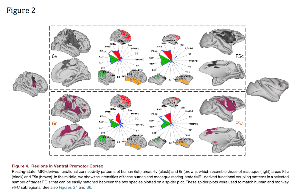
Figure 2 – Click on the image to see a bigger version
Sum Up
- This study offers a fascinating insight into cutting edge research into how human brains and macaque monkey brains compare
- Despite differences in how they were studied, the human and macaque brains demonstrated a striking level of similarity in some areas
- But there were clear differences in others, especially regarding auditory processing
- It is far too simplistic to say that one area is the root of the difference between humans and monkeys
- Improved understanding of how human brains relate to animal brains will hopefully allow us to use less animals in research and find better treatments, quicker
The first rule of blogging: Never pass up the chance to include cute photos of monkeys.
Link
Franz-Xaver Neubert, Rogier B. Mars, Adam G. Thomas, Jerome Sallet, Matthew F.S. Rushworth. Comparison of Human Ventral Frontal Cortex Areas for Cognitive Control and Language with Areas in Monkey Frontal Cortex. Neuron – 30 January 2014. [Abstract]

Are humans like monkeys? MRI scanning suggests similarities and differences that might help future research: D… http://t.co/sUBwcD1haL
New study finds substantial overlap (and key differences) between human and monkey brain function http://t.co/gRwIFNLfZ8
Mental Elf: Are humans like monkeys? MRI scanning suggests similarities and differences that might help future… http://t.co/Z0HVxLJwN9
Human & macaque monkey brains are similar in areas associated with using visual info for motor control & planning http://t.co/gRwIFNLfZ8
New research provides support for the idea that there is not a specific/separate language network in humans http://t.co/gRwIFNLfZ8
Don’t miss: Are humans like monkeys? MRI scanning suggests similarities & differences that might help future research http://t.co/gRwIFNLfZ8
Are humans like monkeys? MRI scanning suggests similarities and differences that might help future research http://t.co/n3bjzOAqNc